Texas A&M veterinarians diagnose cause of mysterious colic
An American Paint Horse was brought to Texas A&M after he began experiencing recurring stints of colic

")
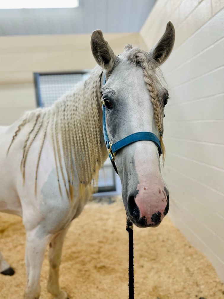
Koche, a 7-year-old American Paint Horse was brought to Texas A&M College of Veterinary Medicine and Biomedical Sciences Large Animal Hospital after his owners noticed he was in pain (Image courtesy of Texas A&M College of Veterinary Medicine and Biomedical Sciences)
Cedrick Harvey brought Koche, a 7-year-old American Paint Horse, to Texas A&M College of Veterinary Medicine and Biomedical Sciences after Koche started to experience flareups of colic. According to Harvey, Koche had been healthy for her entire life up until March 2023 when the family began noticing she was presenting with signs of pain. Koche holds a special place in Harvey's heart because of her family history with the family.
“Her grandmother was an amazing Paint horse named Apache that grew up within our family,” said Harvey, in an organizational release.1 “We really wanted to keep her bloodline alive, so we bred her and got Cody, who was Koche’s father. So, I’ve had Koche since her birth.”
Koche was brought to her local veterinarians to try and determine what was causing her pain, but the doctor was unable to diagnose her but recommended her to the Texas Texas A&M College of Veterinary Medicine and Biomedical Sciences Large Animal Teaching Hospital (LATH). Here, she was able to be treated with more advanced equipment to help diagnose and treat her pain.
“[Horses with colic] do what I call the ‘no-pants dance,’” said Dustin Major, DVM, DACVS (LA), a VMBS clinical assistant professor of large animal surgery.1 “They circle around and posture like they’re about to lay down, but then they never do. Koche was doing that little dance and kicking at her belly, so we knew she was in pain.”
According to the release, colic is a general term used for abdominal pain in horses that can have a wide variety of causes, but each horse experiencing it tends to have the same reaction.1 When Koche arrived at LATH, she was given medication to reduce her pain and she received what is commonly known as the colic workup. Major began a physical exam, checked for gastric reflux with a nasogastric, and a rectal exam to palpate her internal organs. The team determined that she did not have reflux, and all Major could feel on her rectal palpation was some feed in her large intestine, which he explained was nothing out of the ordinary. After the colic workup, the team found nothing that would cause her to show the signs of pain she was presenting with.


The X-ray of the cantaloupe-sized stone found in Koche’s colon and the stone next to a one-inch needle
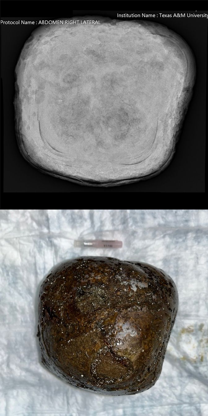
Major then performed an abdominocentesis and an abdominal x-ray, but both provided no answers as well. With no other options, he decided that he needed to go for the final option, colic surgery. Major studied her abdominal organs during the procedure to look for what was causing her pain, and the answer became clear, she had a tone the size of a cantaloupe inside her large colon.1 The stones, known as enteroliths, are formed in the horse's right dorsal colon, the section designed to contract and pack things together. Major explained that this is common in horses in California, but is relatively rare in Texas, which could be why it was tricky to diagnose.
“It’s sort of like making a pearl; the horse ingests a piece of wood, gravel, or something else and then the colon compacts minerals around it, layer upon layer upon layer,” explained Major.1 “The stone then goes down to the transverse colon, where it lodges and backs everything up. The stone can come back out of the transverse colon and then lodge in it again, which is why these horses can be intermittently painful.”
Major was able to remove the 5-pound stone during the colic surgery and after discussing with Harvey, they determined that the enterolith was caused by a high diet of alfalfa combined with high calcium content in the water in Crosby, the town they are from. X-rays were performed on the stone to see if they could determine what was at the center, but the results came back inconclusive.
Her successful treatment was accredited to her local veterinarian, the LATH team, and Harvey’s pushing for someone to help find the source of her pain. Horses that do not receive treatment for their source of colic, including enterolith like Koche, can be at risk of death.
“If you have a horse with colic and it’s not getting better, it’s better to go ahead and be aggressive on the front end,” explained Major.1 “You’ll spend less money and have a better chance of having a successful outcome. Especially from a surgical perspective, the earlier we get into the abdomen, the better. Literally, minutes count.”
Koche stayed at LATH for a week before returning home to her family.
References
- Megan Myers. Texas A&M veterinarians leave no stone unturned in diagnosing horse’s mysterious colic. Texas A&M College of Veterinary Medicine and Biomedical Sciences. Published March 5, 2024. Accessed March 11, 2024. https://vetmed.tamu.edu/news/press-releases/koche/