How to perform a thorough dental exam
Make sure you collect-and record-all the important information during this exam, so the veterinarian can make the correct diagnosis and provide the best therapy.
Technicians are likely to perform preliminary dental examinations to provide initial observations. A thorough evaluation should include performing conscious and anesthetized examinations and charting disease processes, pathology, and anomalies. When performing an examination, be sure to record all findings and bring abnormalities to the veterinarian's attention.

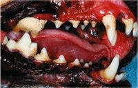

What's wrong with this dog's teeth? Check your answer at dvm360.com/toothquiz.
Conscious examination
An oral examination on a conscious patient is important, but you are often limited to a visual inspection and digital palpation. This initial examination involves more than just the oral cavity. Be sure to palpate the facial bones and zygomatic arch, temporomandibular joint, salivary glands, and lymph nodes. Also be sure to evaluate the dental occlusion, which can be done by gently retracting the lips to look at the soft tissue, the occlusion (bite), and the buccal aspects of the teeth.
Anesthetized examination
Anesthesia is required for both routine dental cleaning and treatment of dental abnormalities or diseases of the oral cavity. Once a patient is anesthetized, you can complete a thorough oral examination. Examine all the structures of the oral cavity, including the oropharynx, lips and cheeks, mucous membranes, hard palate, floor of the mouth, tongue, and teeth. Evaluate the periodontium (gingiva, periodontal ligament, cementum, and alveolar bone) of each tooth.

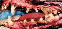
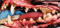
1. A mouth demonstrating mild (A), moderate (B), and heavy (C) calculus.
If patients have large amounts of calculus on their teeth, you may need to remove these deposits to accurately access the periodontium. Calculus (tartar) is mineralized plaque. Using calculus removal forceps is recommended to remove supragingival calculus, but take care to ensure that the gingiva and tooth crown are not damaged.
Record the amount of calculus and plaque observed on the teeth for future reference before cleaning. The amount of calculus and plaque can be recorded as mild, moderate, or heavy (Figure 1). Because plaque is the soft, gelatinous matrix of bacteria and bacterial by-products that lead to gingival irritation and gingivitis, it may be necessary to use a disclosing agent to visualize. Calculus can only be removed by hand scaling or using a power scaler.
When evaluating the periodontium, you will need a periodontal probe, a dental explorer, and a dental mirror. Evaluate the following seven indices for each tooth in each patient.
1—Gingival index
The gingival index is a measurement of gingival health. To assess a patient's gingival health, gently place a periodontal probe into the gingival sulcus (the area between the tooth and free gingiva), and guide the probe around the tooth. Score the assessments of gingival changes by using the following criteria:
- 0 = normal healthy gingiva
- 1 = mild inflammation or redness, no bleeding on probing, possible edema or contour changes
- 2 = moderate inflammation, moderate to severe redness, edema, bleeding on probing
- 3 = severe inflammation or redness, edema, ulceration, spontaneous bleeding
2—Probe depth
Probe depth is a measure of the depth of the periodontal pockets. This measurement increases as a result of inflammation between a tooth and the surrounding tissues. As the inflammation progresses, the gingiva loosens from the tooth. Deep probe depth and the loss of tissue and bone support indicate more severe periodontal disease. Edema and gingival hyperplasia can create a pseudopocket. This increased depth without the loss of attachment can progress into periodontal disease unless treated.
To measure probe depth, gently place a periodontal probe with millimeter markings between the free gingiva and the tooth surface, and carefully advance it until you feel soft tissue resistance. The tip of the probe should be parallel to the long axis of the tooth. Record the probe depth as the distance in millimeters from the free gingival margin to the bottom of the pocket. You may glide or walk the probe along the tooth to measure the varying depths. A normal gingival sulcus depth is 1 to 3 mm in dogs and 0.5 to 1 mm in cats. Record measurements in excess of these values in the appropriate location on the dental chart.
3—Gingival recession
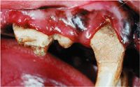
Technicians also need to use the periodontal probe to measure the gingival recession or root exposure, which is the distance from the cemento-enamel junction to the margin of the free gingiva. At sites with gingival recession or root exposure (Figure 2), the probe depth may be normal despite the loss of alveolar bone. Note areas of gingival recession on the dental chart.

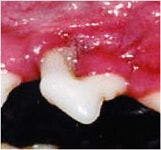
2. A tooth demonstrating gingival recession and Stage 3 furcation.
4—Periodontal attachment level
This measurement is similar to probe depth except that the pocket depth is measured from the base or apex of the pocket to the cemento-enamel junction. This is a more accurate assessment of tissue loss from periodontitis than probe depth. The periodontal attachment level can be measured directly, or it can be calculated as the sum of probe depth and gingival recession.
5—Furcation exposure
In multirooted teeth, the area where the roots join the crown is referred to as the furcation. The furcation index measures the loss of bone support in multirooted teeth. The bone loss caused by periodontal disease often affects this area early in the disease process.
Place a periodontal probe about perpendicular to the long axis of the tooth, and slide it along the free marginal groove to the furcation site. Record the involvement as stage 0 to 3 using the following criteria:
- 0 = no loss of bone support
- 1 = horizontal loss of supporting tissues not exceeding one-third of the width of the tooth
- 2 = horizontal loss of supporting tissues exceeding one-third of the width of the tooth but not encompassing the total width of the furcation area
- 3 = complete horizontal loss of supporting tissue (Figure 2)
Be sure to note a furcation index of 0 to 3 on the dental chart.
6—Tooth mobility
The amount of tooth movement indicates the level of bone support loss. To measure tooth mobility, place the length of the periodontal probe on the buccal surface of the tooth's crown, and apply gentle pressure. Use the following criteria to assign a numerical score (stage):
- 0 = no mobility
- 1 = perceptible mobility, but less than 1 mm buccolingually
- 2 = definite mobility between 1 and 2 mm
- 3 = gross mobility exceeding 2 mm buccolingually or vertical mobility


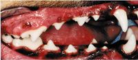
3. Stage 1 periodontal disease. Note the mild inflammation on the maxillary canine.
Note a mobility score of 0 to 3 (M0, M1, M2, M3) on the dental chart.
7—Stage of periodontal disease
Noting the severity of periodontal disease can be used to help the veterinarian determine the appropriate therapies, and it also needs to be recorded so that disease progression can be monitored. The stage of periodontal disease is determined either radiographically or by measuring clinical attachment level and assigning a severity score as follows:
- Normal = no gingival inflammation
- Stage 1 (Figure 3) = gingivitis only, without attachment loss
- Stage 2 (Figure 4) = less than 25 percent attachment loss, stage 1 furcation (multirooted teeth)
- Stage 3 (Figure 5) = 25 percent to 50 percent attachment loss, stage 2 furcation (multirooted teeth)
- Stage 4 (Figure 6) = more than 50 percent attachment loss, stage 3 furcation (multirooted teeth)


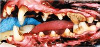
4. Stage 2 periodontal disease.
These tips for tracking dental abnormalities may help catch potentially serious problems early and enhance and extend pets' lives.

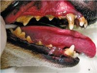

5. Stage 3 periodontal disease.

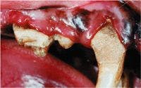
6. Stage 4 periodontal disease. Note the gingival recession and furcation on the maxillary second premolar.
Mary Berg, BS, RVT, RLATG, VTS (dentistry), is the practice manager and dental technician specialist at Gentle Care Animal Hospital in Lawrence, Kan. She is also president of MLB Consulting, a veterinary dental consulting company.
RECOMMENDED READING
1. Logan EI, Boyce EN. Oral health assessment in dogs: parameters and methods. J Vet Dent 1994;11(2):58-63.
2. Harvey CE, Emily PP. Periodontal disease. In: Harvey CE, Emily PP, eds. Small animal dentistry. St. Louis, Mo: Mosby, 1993.
3. Logan EI, Finney O, Hefferren JJ. Effects of a dental food on plaque accumulation and gingival health in dogs. J Vet Dent 2002;19(1):15-18.
4. Holmstrom SE. Veterinary dentistry for the technician and office staff. Philadelphia, Pa: WB Saunders Co., 2000.
5. Gorrel C. Veterinary dentistry for the general practitioner. Philadelphia, Pa: Saunders, 2004.
6. Lobprise H. Five-minute consult clinical companion: small animal dentistry. Ames, Iowa: Blackwell, 2007.