How to ace autoimmune skin disease diagnostics
Cytology slip-ups to avoid, biopsy tips to try and why you need to perform complete blood count and chemistry tests (even when the veterinary patients spirits and energy level are high).
Your patient's body is attacking itself. Before you can develop your plan of attack, you'll need to perform these tests and perform them well to determine what you're fighting.
Avoid these cytology slip-ups
A cytology test is simple to perform, but there are many common pitfalls to avoid:
First, you must consider whether it is more appropriate to use a glass slide or a piece of clear packing tape to collect the sample. Slides can be easily pressed onto moist or gooey lesions and can also be used to lift the edge of a crust to maneuver underneath. Tape is most helpful when collecting samples from dry lesions and from areas too small for the glass slide.
Next, you need to stain your sample. Tape does not need to be dipped in a fixative and should not be heated.
Lastly, your ability to evaluate cytology will improve with time, but you can set yourself up for better results by upgrading your dilapidated microscrope to a newer model.
6 tips for a better biopsy
A biopsy is the ultimate dermatology test, but it won't always give you a straight answer. Here are six tips for better results:
- Collect multiple samples. In most cases, aim for four to six pieces of tissue.
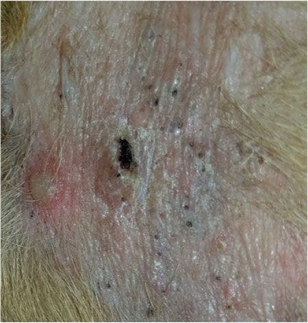
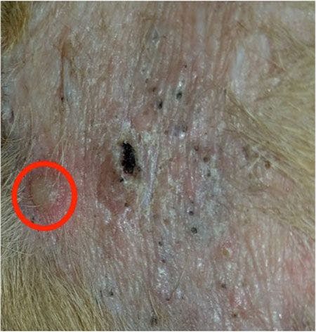
- Center your biopsy punch on the lesion (see page 2 for examples). Do not send normal tissue to the pathologist. If you send in the margin of a lesion and include normal tissue, there is a risk that the lab technician will not “cut in” the diseased tissue for examination.
- If you see an infection on cytology (see page 3 for examples), consider resolving it before collecting biopsy specimens. Infection can obscure the primary disease and make the pathologist's job much more difficult.
- Send a thorough history along with your samples.
- Include clinical photographs with the samples when possible (see page 4 for examples).
- Who you send your samples to could make the difference between the right answer, the wrong answer or no answer at all, so send your tissue samples to a dermatopathologist.
The case for complete blood count and serum chemistry profiles
These tests may not seem particularly related to the skin disaster on your exam table (especially when the dog is happy and energetic), but they are important for ruling out underlying metabolic problems that could be causing the disease. The tests are typically used as a guide when determining medications for treatment.
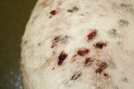
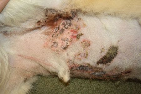
Examples of where to place the biopsy punch


A dog with sterile nodular panniculitis. (All images courtesy of Dr. Darin Dell)

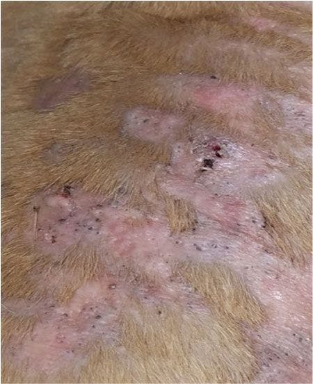


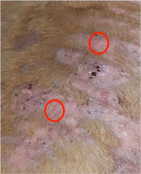
Another view of sterile nodular panniculitis on the same dog.
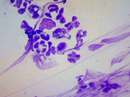
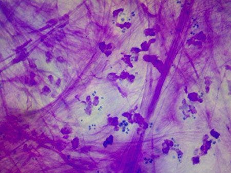
Examples of infection seen on cytology

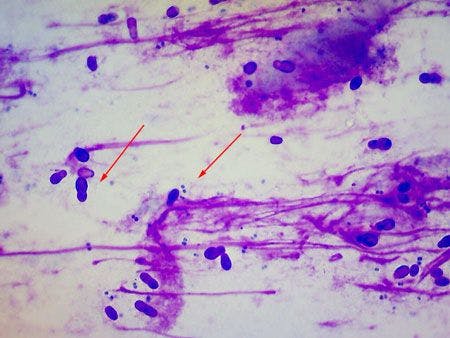

Fireworks yeast and cocci (All images courtesy of Dr. Darin Dell)
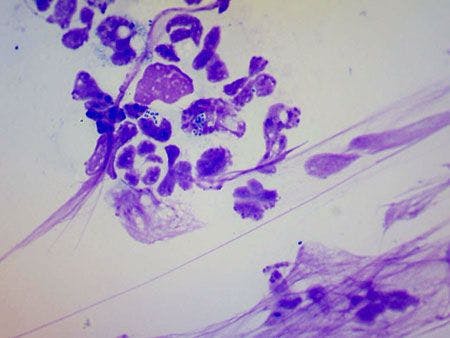
Neutrophil and cocci

Degenerative neutrophil and cocci
Examples of photos sent with skin samples


Nodular panniculitis (All images courtesy of Dr. Darin Dell)

Cutaneous adverse drug reaction
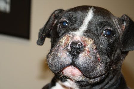

Juvenile cellulitis (puppy strangles)
Dr. Darin Dell spent six years in general practice and two years in emergency medicine before becoming a diplomate of the American College of Veterinary Dermatology in 2012. He is currently on staff at Animal Dermatology Clinic in Indianapolis. Dr. Dell's hobbies include woodworking and mountain biking.