Diagnostic dental radiographs: A concise how-to
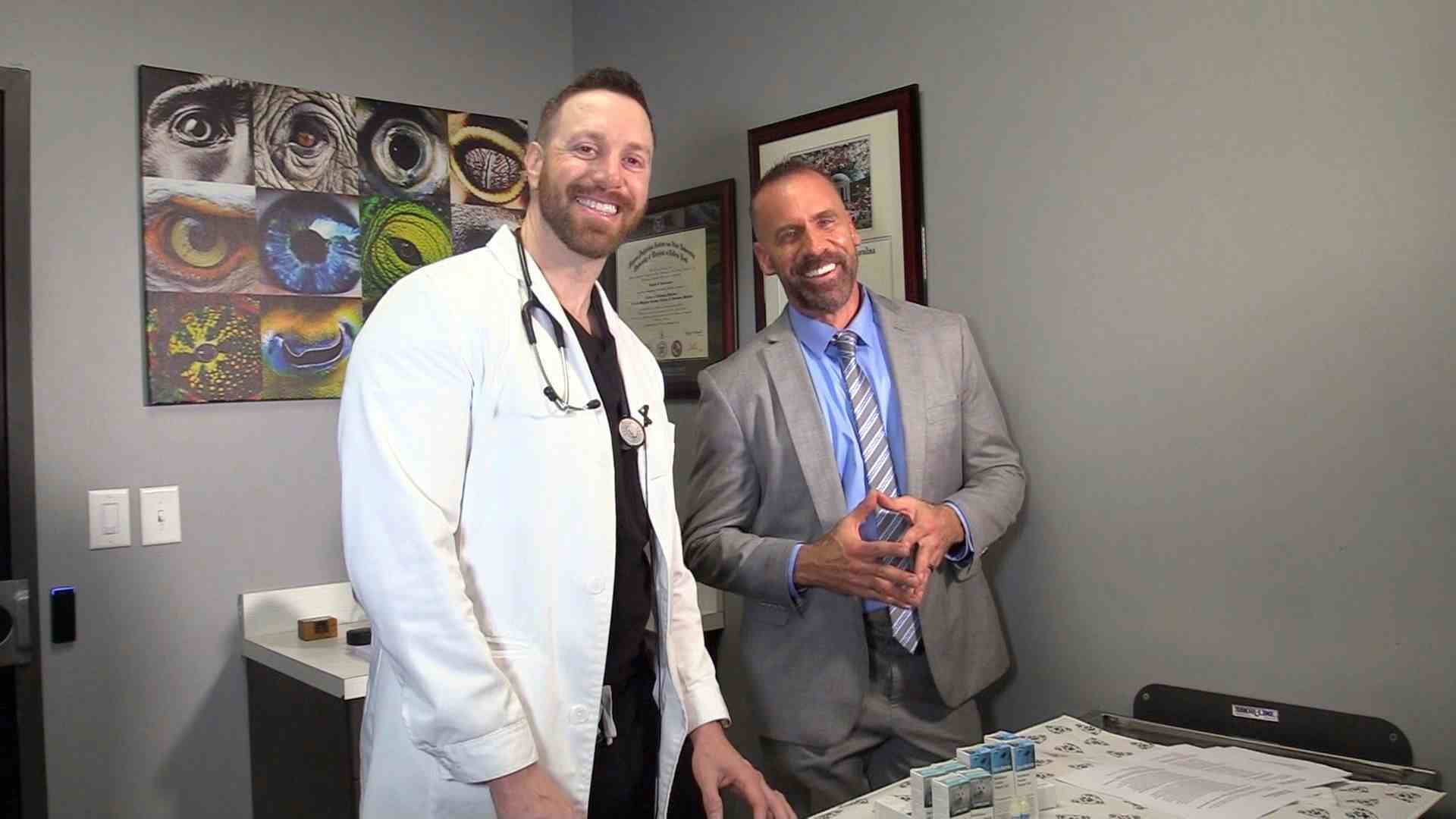
Mary Berg, RVT, RLATG, VTS (Dentistry), demonstrates her preferred method of obtaining these images.
Mary Berg, RVT, RLATG, VTS (Dentistry), has been teaching veterinary technicians how to take dental radiographs for more than 20 years, and here she shares her practiced techniques for each tooth. What makes it a diagnostic radiograph? The image must include 2- or 3-mm of bone around the apex of the root and the level of the alveolar bone, Berg says.
Berg starts by placing the patient in sternal recumbency, and the entire maxilla can be imaged in that position. Prop a towel under the patient's chin to position the maxilla parallel with the table. Place the digital sensor parallel to the table and against the teeth for each maxillary shot to ensure imaging the root and the bone surrounding each tooth.
One tip for sensor stabilization: If the sensor starts to tip a bit, place a piece of gauze between the sensor and the palate to keep the sensor level.
Berg's simplified positioning methods eliminate the need for bisecting angle calculations. Most dental radiography systems already have the angles marked on the unit.
Let's dive in to canine dental radiographs …
To isolate the first and second maxillary molars, Berg places the caudal edge of the digital sensor at the caudal edge of the second maxillary molar and the X-ray tube at 60 degrees and shoots “high and through the eye” and just a bit caudally.
To radiograph the fourth premolar, Berg shifts the sensor forward (rostrally) a bit so it sits right under the tooth, then adjusts the X-ray tube to about 50 degrees and shoots straight at the fourth maxillary premolar.
She again moves the sensor rostrally to image the maxillary premolars. She adjusts the tube to 45 degrees and still aims high. “Always remember you're shooting at the root, not at the crown of the tooth,” she says.
The maxillary canine tooth can be difficult to image because 2/3 of the tooth angles caudally within the maxilla under the nasal area of the dog. To help with positioning, Berg makes an L shape with her index finger on the midline of the dog's head pointed toward the nose and her thumb gently draped over the dog's eye. She sets the X-ray tube at 70 degrees and points it into the corner where her thumb joins her hand. (See this at 3:51), while aiming at the apex of the canine tooth.
For the maxillary incisors, Berg says she can usually radiograph all six in one image. Move the sensor rostrally so the incisors are positioned at its leading edge. Then angle the tube at 45 degrees and move and center it in front of the incisors-still aiming somewhat high at the teeth to image the roots.
Berg then repeats the process on the opposite side of the maxilla.
Now on to the mandible …
Position the dog in dorsal recumbency and place a towel under its neck to keep the mandible parallel to the table. She starts by radiographing the third molar to the fourth premolar with parallel shots, where the digital sensor is parallel to the mandible.
Place the sensor at the back of the mouth so you can just see the sensor above the crowns of the teeth (use gauze as needed to help with sensor positioning) and place the tube head at 90 degrees or perpendicular to the sensor to obtain the third to first molar shots, and then move the sensor forward (rostrally) and just slightly deeper into the sulcus between the jaw and the tongue, radiograph the fourth premolar and the first molar.
For the rest of the mandibular teeth, remember “don't fight the tongue,” it's ok to place the tongue between the teeth and the sensor, Berg says.
So, for the first, second and third premolars, place the sensor parallel to the table and then position the tube head at 60 degrees and shoot following the line of the jaw.
For the mandibular canines and incisors, it's possible to get them into one shot, but to radiograph the canine tooth alone, place the sensor with the leading edge at the canine tooth, position the tube head at 70 degrees and shoot at the apex of the canine tooth root at about the level of the second premolar.
Then position the sensor rostrally, set the tube angle at 45 degrees and radiograph the incisors.
To finish, continue along the jaw in the same way to radiograph the teeth on the opposite side of the mandible.
Time for our feline friends …
Berg says that it used to be really hard to take dental radiographs in cats, but digital radiography has made it much easier. She starts with cats in sternal recumbency for the maxillary teeth and dorsal recumbency for the mandibular teeth, just as she does with dogs.
One of the coolest things about digital radiography with cats is that Berg can place the sensor and image all of the maxillary teeth by moving the sensor little, if at all.
Another cool thing about digital radiography is that you can usually shoot though the zygomatic arch. With film radiography, the arch obscures the teeth views, but that's not the case with digital imaging.
To take images of the second, third and fourth premolars and the first molar, place the sensor in the patient's mouth and parallel to the table, and then set the tube head to 37 degrees. You still want to position the tube head “high and through the eye” and along the line of the jaw, Berg says.
Then, without moving the sensor, Berg sets the tube angle to 50 degrees and moves it to aim at the medial canthus of the cat's eye, allowing her to isolate the canine tooth.
Next, she moves the tube head straight on from the front of the cat's face at 45 degrees and images the incisors. Berg notes that the maxillary canine and incisor teeth in cats cannot be radiographed in one view because the canine teeth will be superimposed over the cheek teeth.
Follow this pattern around the other side of the head to radiograph the maxillary teeth on the opposite side.
For the feline mandible …
With the cat in dorsal recumbency, again with a towel under the neck or shoulders to keep the mandible parallel to the table, place the sensor flat in the mouth. Remember, “Don't fight the tongue. You can place the tongue between the sensor and the teeth,” Berg says.
Then set the tube head to 60 degrees and place it following the line of the jaw and you'll be able to radiograph the mandibular molars and premolars.
Now you're already in place to radiograph the canines and incisors in one view. Simply leave the tube angled at 60 degrees and shift the tube's position to straight in front of those teeth.
Finally, leave the tube angled at 60 degrees and shift the tube's position to the opposite side of the mandible to radiograph the opposite cheek teeth.
And there you have it. Diagnostic dental radiographs for canine and feline patients-without the dreaded bisecting angle calculations!


")