Dental technicians: Filling a vital role in veterinary medicine
Specialty training pays off.
Small-animal veterinary dentistry is filled with people committed to bringing the concept of oral health to the forefront of the veterinary profession. The veterinary technician plays a pivotal role in the execution of the many facets of high-quality dentistry. The key is a solid foundation of education and knowledge, along with the ability to apply the techniques learned with expertise and confidence.
Most of us have entered the veterinary field because we love animals. And you can put your skills to work by pursuing the source of the No. 1 disease in veterinary medicine: periodontal disease. When we educate clients about effective dental care, we promote a lifetime of oral health and well-being for their pets. As a result, the pain and suffering that is so notable with dental disease can be prevented or addressed and alleviated. The reward is great for technicians as professionals. And for pets, it improves their level of comfort and quality of life.
This article examines three cases from the files of the Academy of Veterinary Dental Technicians (AVDT) that represent different aspects of oral disease seen in small-animal veterinary dentistry. These cases offer insight into the importance of recognizing and implementing good oral health for all of your patients.
CASE 1: Painful gingivitis and periodontitis in a kitten
Juvenile gingivitis or periodontitis can be a painful condition for a young animal and disheartening to the pet's owner. Clinical signs include extensive inflammation of the gingival tissues, oral pain, hesitancy to eat and groom, and bleeding of the gums, and it can lead to periodontal disease and tooth loss.
Tianna, a 6-month-old female kitten, was presented for a physical examination and vaccinations. The owner reported she would pull away when he tried to pet her and would not allow him to handle her mouth to any degree. He reported some bleeding from her mouth. The owner had already been through extensive oral therapy with his older cat and was aware of the signs the kitten was showing.
On oral examination, it was obvious that Tianna's gingiva was inflamed and already showing some attachment loss along several teeth. The owner was advised of the importance of the mouth being kept as clean as possible, which ideally meant brushing the teeth to remove the plaque on the tooth surfaces. But this process would be painful for Tianna and difficult for the owner to accomplish. So Tianna was scheduled for an oral assessment under general anesthesia to help choose the most beneficial treatment.

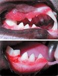

Figure 1 & 2. Generalized gingivitis is evident on oral examination.
Tianna underwent a preanesthetic blood panel, had an intravenous catheter placed, and was maintained under general anesthesia. An oral examination revealed severe gingival inflammation with periodontitis (Figures 1 and 2). A full-mouth series of digital radiographs was obtained, which revealed several teeth with marked vertical bone loss and mobility (Figure 3). A supernumerary left mandibular premolar (tooth 308) was also involved (Figure 4).


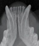
Figure 3. An intraoral radiograph revealing horizontal bone loss on the mandibular incisors.
It was recommended to clean the teeth and extract any teeth showing evidence of periodontal disease. All of the mandibular incisors were removed, along with the supernumerary tooth 308 and the left mandibular molar (tooth 309). Extensive gingival hyperplasia was also present, so gingivectomy with a carbon dioxide laser was performed where needed (Figure 5). Tianna's recovery was uneventful.

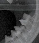
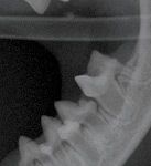
Figure 4. An intraoral radiograph revealing a supernumerary tooth (308) as well as vertical bone loss on tooth 309.
Tianna was released that evening with instructions for follow-up care, including using an oral rinse and continuing a regimen of antibiotics and pain medication that had been started before the oral procedure.

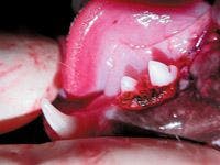

Figure 5. The gingivectomy site on teeth 307 and 308 as well as the extraction site of teeth 308 and 309.
Her first recheck appointment in one week showed healing of the extraction sites and gingival tissues along with a noticeable reduction of gingival inflammation. The owner was advised to return for another oral examination in three months—and sooner if needed.
Juvenile gingivitis or periodontitis requires an owner's conscientious care and commitment to ensure continued treatment, since the condition is not easily resolved. It is hoped that Tianna will continue to improve and be able to live her life without the constant pain she endured early on. Oral examinations throughout the year along with any therapy needed will be important to Tianna's long-term oral health.
CASE 2: An unusual malocclusion in a cat
At Community Animal Hospital in Reno, Nev., a licensed technician is assigned to each client for a dental consult, which includes oral examination, discussion of the oral findings, and home-care recommendations and instructions. As this case illustrates, it is imperative that each patient receive a thorough, awake oral examination by a qualified technician or doctor. This person must have the ability to recognize oral pathology and abnormalities when present. There is no reason that any animal should suffer with pain from an unrecognized oral condition.

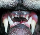
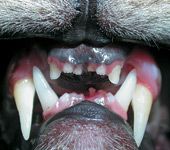
Figure 6. The palatal trauma is evident around teeth 304 and 204.
In this case, Catarina, a 3-year-old spayed female cat, was presented as a new patient for her annual physical examination and vaccinations. When the assigned technician was conducting the dental consult, the client mentioned that Catarina had an overbite, which had been noted by a previous veterinarian early in Catarina's life. A class 2 malocclusion is not a common finding in cats and made the technician question why this cat developed this type of abnormal bite.


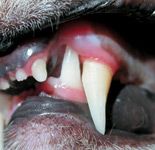
Figure 7. Tooth 304 is causing dental interlock.
On oral examination, it was discovered that Catarina had a base narrow, left mandibular canine (tooth 304) causing deep tissue trauma to the palatal aspect of the left maxillary canine (tooth 204) (Figure 6). More than one-fourth of the crown was buried into the palate, causing severe discomfort and a dental interlock (Figure 7). This condition had caused the maxilla to extend beyond the mandible during the growth period, with the mandibular canine tooth acting as a blocking agent to impede the natural growth sequence of the mandible. The incisors were crowded and skewed because of the abnormal occlusion.


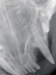
Figure 8. An intraoral radiograph revealing palatal bone loss due to tooth 204.
When relating the cat's history, the owner revealed that Catarina had always shown some difficulty chewing her food. She would drop it and then rapidly eat what she had and swallow it. This painful malocclusion had gone unnoticed, and Catarina had suffered with her canine tooth probing into her hard palate for most of her life.
The treatment plan included performing a prophylaxis, obtaining full-mouth radiographs, and extracting tooth 304. Catarina's procedure was scheduled. A blood panel showed no abnormalities.

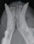

Figure 9. An intraoral radiograph revealing greater than 50 percent bone loss on teeth 301, 302, and 303.
Catarina returned the next week for her dental procedure. After an intravenous catheter was placed and premedications were administered, Catarina was anesthetized. Radiographs showed marked loss of palatal bony structure to the left maxillary canine (tooth 204) because of the constant contact and pressure elicited by the mandibular base narrow canine (Figure 8). The left mandibular incisors (teeth 301, 302, and 303) all had extensive bone loss (Figure 9) with level 3 mobility. No other pathology was found on the remaining radiographs or anesthetized oral examination.
A mental nerve block was given using a mix of bupivacaine and lidocaine, and the prophylaxis was performed. Tooth 304 was surgically extracted, along with simple extraction of teeth 301, 302, and 303. A synthetic bone grafting material was placed in the extraction site of tooth 304. The sites were closed with 5.0 Monocryl (Ethicon).
Catarina recovered successfully from anesthesia, and she was released to her owner by late afternoon. Catarina was sent home with antibiotics and oral buprenorphine for pain management, along with instructions for home care, including soft food for the following two weeks.
Catarina returned in one week for a recheck of her extraction sites. Catarina's recovery period was smooth and rapid. She could finally eat without a struggle. The owner expressed amazement at the cat's happy demeanor and contentment.
CASE 3: Root canal vs. extraction
Tooth preservation is the primary goal in veterinary dentistry, so when an animal has a fractured or nonvital tooth, a decision has to be made. In Hilda's case, the owner wanted to do whatever was necessary to save her tooth.

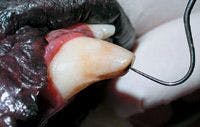
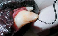
Figure 10. Hildaâs fractured tooth (304).
Hilda is a 6-year-old German shepherd that was presented with a fractured mandibular left canine (tooth 304) with vital pulp exposure (Figure 10). Discussion during the initial examination centered on the owner's ability to modify Hilda's destructive chewing habits, which had caused the fracture in the first place. Hilda's destructive habits consisted of aggressive, constant chewing on hard objects and surfaces, including hard toys with no give or flexibility. Behavior modification would be crucial to the successful long-term outcome of root canal therapy. If the destructive behavior continued, extraction of the tooth might be a better alternative. Hilda's owner was committed to curbing her dog's habits and chose root canal therapy.

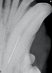

Figure 11. An intraoral radiograph showing the fractured drill shaft in the canal.
The procedure was performed with an engine-driven, rotary instrumentation system for performing root canals. It improves the treatment technique, providing faster, more efficient performance of the root canal procedure. In contrast to traditional hand filing techniques, these instruments allow for more efficient cutting and shaping of the canal walls while conserving unaffected dental tissues.
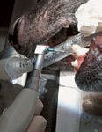

Figure 12. The coronal access site.
Initial access was made, and the necrotic pulp contents were removed with a No. 15 K-file. During enlargement of the coronal third of the canal with a drill, the tip became bound, and the shaft fractured off (Figure 11). With persistence, EDTA flushes, and further enlargement of the canal, the dog's head was lowered, its jaw was tapped three or four times, and the drill shaft appeared at the access. It was removed immediately and the procedure continued.


Figure 13. The placement of the gutta-percha plug.
The working length was determined, and nickel-titanium drills were used to reach the apical access (Figure 12). The master apical rotary was then noted and confirmed. The mid-canal was addressed, filed, and cleaned. A sealer was applied with a spiral filler, and a gutta-percha plug was applied to the apical position (Figure 13). The canal was then backfilled for complete obturation (Figures 14 & 15). The original access opening was cleared with a No. 330 pear-shaped bur, and an intermediate restorative layer of glass ionomer was applied and light-cured. The restoration was completed with a composite restorative material and a sealer applied to prevent leakage around the margins of the composite seal (Figure 16).

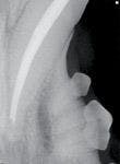
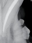
Figure 14. An intraoral radiograph revealing obturation to the apical access.
Hilda has returned for periodic oral and radiographic examinations of her canine tooth root canal, and, to date, the restoration is intact and the root canal therapy remains successful. Although extraction is an acceptable option—and in some cases the better option—preserving the function of a major tooth in this canine's oral dentition proved to be the best option.

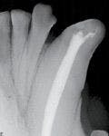
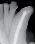
Figure 15. An intraoral radiograph revealing coronal obturation.
Conclusion
Technician specialties give technicians the ability to raise their level of expertise and proudly claim the title of technician specialists in their chosen field. To learn how to pursue a veterinary technician specialty in dentistry, visit the AVDT website at avdt.us. Every veterinary clinic should make an effort to raise the standards of their team's dental-related education to greatly increase revenue and, more important, to give patients the best care possible. This is an ever-advancing field, and more and more clinicians and pet owners are demanding a higher level of dental care from veterinary technical personnel.


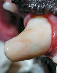
Figure 16. The completed restoration of the root canal site.
Nancy Smith LVT, VTS (Dentistry), has been in small-animal practice for more than 20 years in the Carson City/Reno area of Nevada, concentrating her interests on small-animal dentistry. She continues to promote the importance of good oral care and preventive dentistry through educational lectures, labs, and an emphasis on client education.