Dental corner: Correcting a congenital cleft palate in your veterinary practice
This veterinary hospital team works together to rid a Rhodesian ridgeback of a congenital cleft palate.
Mika, a 6-month-old, female Rhodesian ridgeback, was presented to the Dentistry and Oral Surgery Service at the University of Pennsylvania for evaluation of a cleft palate. Mika's defect was noted at birth. The breeder tube fed the puppy for the first eight weeks, and then Mika was placed in a new home. She suffered from several episodes of aspiration pneumonia, which responded to antibiotics. The results of a preoperative laboratory work-up were normal, and she was deemed a good surgical candidate.
Surgery
In order to ensure the flap would be big enough to cover the defect, Mika had surgery the day of her initial exam to extract three premolars on her right maxilla. Four weeks later, she returned for palatal surgery.


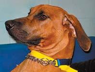
Photo 1: Mika at her initial exam. (PHOTOS COURTESY OF THE UNIVERSITY OF PENNSYLVANIA)
Mika was anesthetized, and her bilateral secondary hard and soft cleft palate defect was surgically corrected. The mucoperiosteum of the hard palate was incised to create a releasing or elevating flap on the right side of the palate. This flap was folded over so that the connective tissue surfaces were in contact and sutured closed. This technique allowed for coverage of the midline defect. Note the difference in the rugae folds of the palate in Figure 3.
Recovery
An Elizabethan collar was placed, and Mika was sent home with antibiotics and analgesics and instructions to feed soft food only for two to four weeks. No toys or chewing were allowed.

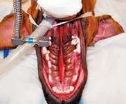

Photo 2: The missing teeth and the healed extraction site are visible. The palatal defect extends the length of the hard and soft palate. (PHOTOS COURTESY OF THE UNIVERSITY OF PENNSYLVANIA)
Mika recovered well. At her recheck exam four weeks later, a small area of tissue on the rostral maxilla, just caudal to the incisive papilla, had retracted and opened. The owner was given the option for another surgical procedure to close the small defect. The owner elected to postpone another surgery.
At last report, Mika is a happy, healthy adult, and the small open area on the rostral palate does not seem to be causing any issues at this time.
Discussion
Palatal defects may be acquired—due to trauma or severe periodontal disease—or congenital, as in this case. Congenital palatal defects may have a hereditary component or may result from metabolic disorders or vitamin imbalances in the dam or exposure of the dam to teratogenic chemicals or drugs. These defects are classified as either primary (involving the rostral region on the lateral aspects of the palate) or secondary (affecting the areas of the caudal palate along the midline). Affected neonates typically have difficulty nursing and swallowing, often regurgitating and aspirating. This can progress to pneumonia and eventually death.


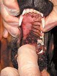
Photo 3: The palate surgery site. The left side is normal tissue with rugae folds. The right side is smooth, because it is the underside of the palatal tissue that was folded over to create a flap and repair the defect. The rugae folds are now on the inside. (PHOTOS COURTESY OF THE UNIVERSITY OF PENNSYLVANIA)
Surgical intervention is almost always necessary. Most oral surgeons will wait until a patient is 4 to 6 months old, as in this case, to ensure that the palate is growing properly and that the tissue has sufficient strength to withstand the surgical repair.
A dental technician must know the anatomy of the palate, what causes the defects and how they can be repaired. If technicians and doctors aren't aware of surgical options, pets with palatal defects might be euthanized.
Technicians may need to teach clients how to tube feed puppies and kittens until they are able to have surgery.
Dental technicians assist during surgery, handing instruments, retracting tissue and cutting suture. After surgery, they monitor the pet's recovery.
At checkout, a dental technician will discuss possible complications with the owners as well as an appropriate feeding schedule and medication use.
Patricia March, CVT, VTS (Dentistry), is a dental technician at Animal Dental Center in Baltimore, Md., and the past president of the Academy of Veterinary Dental Technicians.