Dental Corner: A Boxer with Gingival Hyperplasia
A veterinary team tames excessive gingival tissue in a boxer.
Rocky, an 8-year-old, neutered, male boxer, was presented at the University of Pennsylvania's Dentistry and Oral Surgery service for evaluation of gingival masses. It was difficult to locate normal gingival tissue in his mouth. Multiple oral masses were visible, and the crowns were almost completely covered by the excessive tissue (gingival enlargement).

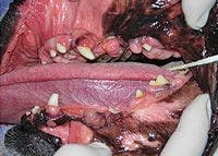

Photo 1: The left side of Rockyâs mouth showed all the excessive gingiva and the crowns barely seen due to the overgrowth.
Complete blood count and serum chemistry profile results were normal as were preoperative chest radiographs. Rocky was anesthetized, and intraoral radiographs were taken to evaluate the teeth and jawbones.
The veterinarian noted class 3 malocclusion, or mandibular distoclusion, in which the mandible is longer and wider than the maxilla. Rocky was missing several teeth, and a few teeth were affected by periodontal disease.
Treatment
The oral masses were measured, evaluated, and surgically removed with a No. 15 surgical blade, followed by a 12-fluted bur on a high-speed handpiece to contour the gingiva. Rocky's remaining teeth were ultrasonically scaled and polished. Four diseased premolars were extracted. Areas of periodontal pocketing were treated with gingival curettage and a locally applied antimicrobial gel.
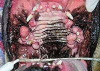

Photo 2: Rockyâs maxilla and palate had excess tissue and several round masses.
Diagnostic testing and follow-up care
Biopsy samples of several oral masses were obtained. Most of the masses were diagnosed as gingival hyperplasia, but two of them were fibromas.


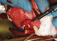
Photo 3: The enlarged tissue was excised by using a scalpel blade.
Gingival hyperplasia is a common, benign condition in which the gingiva grows at an abnormal rate and can cover the crowns of the teeth, creating pseudopockets that trap debris and bacteria and affect periodontal health. The affected gingiva must be surgically removed, and the overgrowth will likely recur. Certain medications, such as cyclosporine,1 have been linked to increased formation of excessive gingival tissue. However, the recommended treatment is the same.
Fibromas are benign oral masses, often found on the gingiva, that should be removed and sent for histopathologic diagnosis since they can mimic malignant gingival tumors.

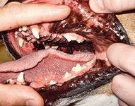
Photo 4: Rocky was healing well at his two-week recheck.
Rocky was sent home with antibiotics, analgesics, and instructions to feed soft food only for two weeks. At his recheck appointment, his gingiva was healing well, and Rocky seemed comfortable.
Patricia March, CVT, VTS (Dentistry), is a dental technician at Animal Dental Center in Baltimore, Md., and the past president of the Academy of Veterinary Dental Technicians.
Reference
1. Beckman B. Gingival hyperplasia. NAVC Clin Brief 2010:11-14.